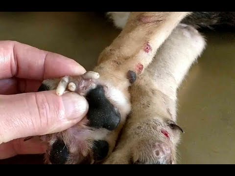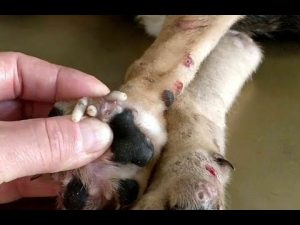

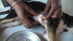
The mode of infection by the Cayor Worm. Doctors Rodhain and Bequaert conclude, from their observations in the Congo Free State, that Cordylobia anthropophaga lays its eggs on the ground. The larvae, known generally as Cayor Worms, crawl over the soil until they come in contact with a mammal, penetrate the skin and lie in the subcutaneous tissue, causing the formation of tumors. On reaching full growth, the larvae leave the host, fall to the ground, bury themselves and then pupate. This fly is said to be the most common cause of human or animal myiasis in tropical Africa, from Senegal to Natal. In the region of Lower Katanga where these investigations were made, dogs appeared to be the principal hosts, although Cordylobia larvae were found also in guinea-pigs, a monkey, and two humans. The larvae are always localized on those parts of the hosts which come in immediate contact with the soil.”
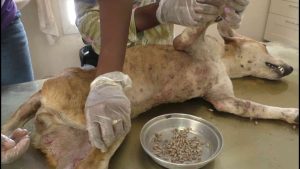
The larvae of the tumbu fly, Cordylobia anthropophaga, were first described in Senegal in 1862, and Blanchard first described the adult and gave it its name in 1872. In 1903, Grünberg placed the tumbu fly in a new genus, Cordylobia.
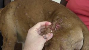
Female tumbu flies deposit around 300 eggs in sandy soil, often contaminated with feces. The hatched larvae can remain viable in the soil for 9–15 days, when they need to find a host to continue developing.[4] If a larva finds a host, it penetrates the skin and takes 8–12 days developing through three larval stages before it reaches the prepupal stage. It then leaves the host, drops to the ground, buries itself, and pupates. It then becomes an adult fly able to reproduce and begin the cycle all over again.
Clinical presentation in humans
[edit]
Successful penetrations in humans results in furuncular (boil-like) myiasis, typically on the backs of arms or about the waist, lower back, or buttocks.[6]
C. anthropophaga rarely causes severe problems, and mainly causes cutaneous myiasis. Geary et al. describe the presentation of cutaneous myiasis caused by the tumbu fly: “At the site of penetration, a red papule forms and gradually enlarges. At first the host may experience only intermittent, slight itching, but pain develops and increases in frequency and intensity as the lesions develop into a furuncle. The furuncle‘s aperture opens, permitting fluids containing blood and waste products of the maggot to drain.”[5]
